

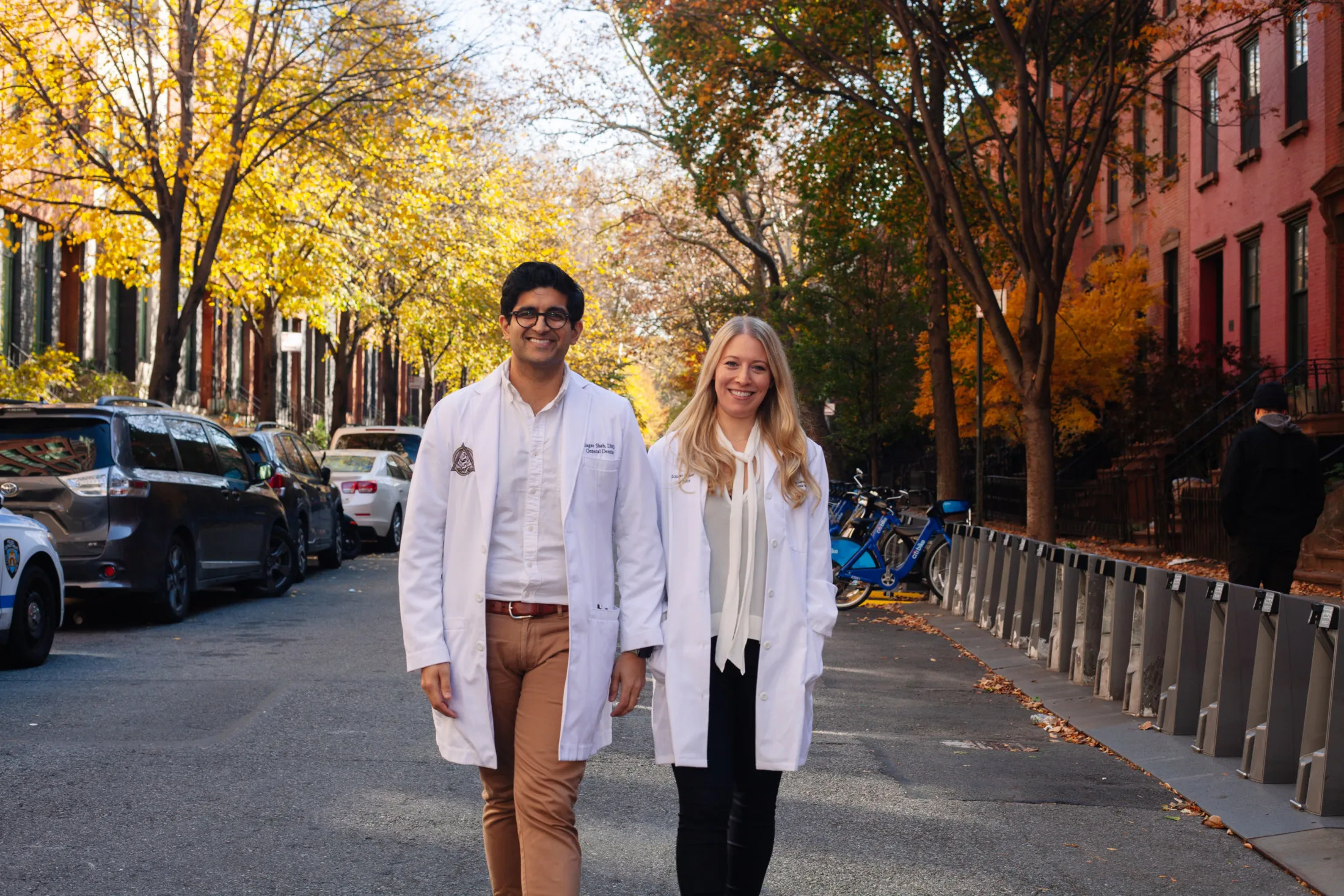
Downtown Dental Office Tour & Technology
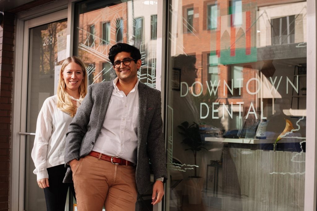


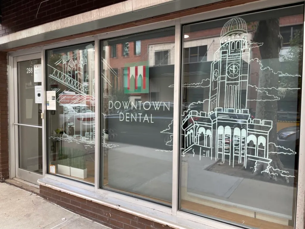


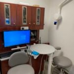

Patient Care & Comfort in Downtown Brooklyn
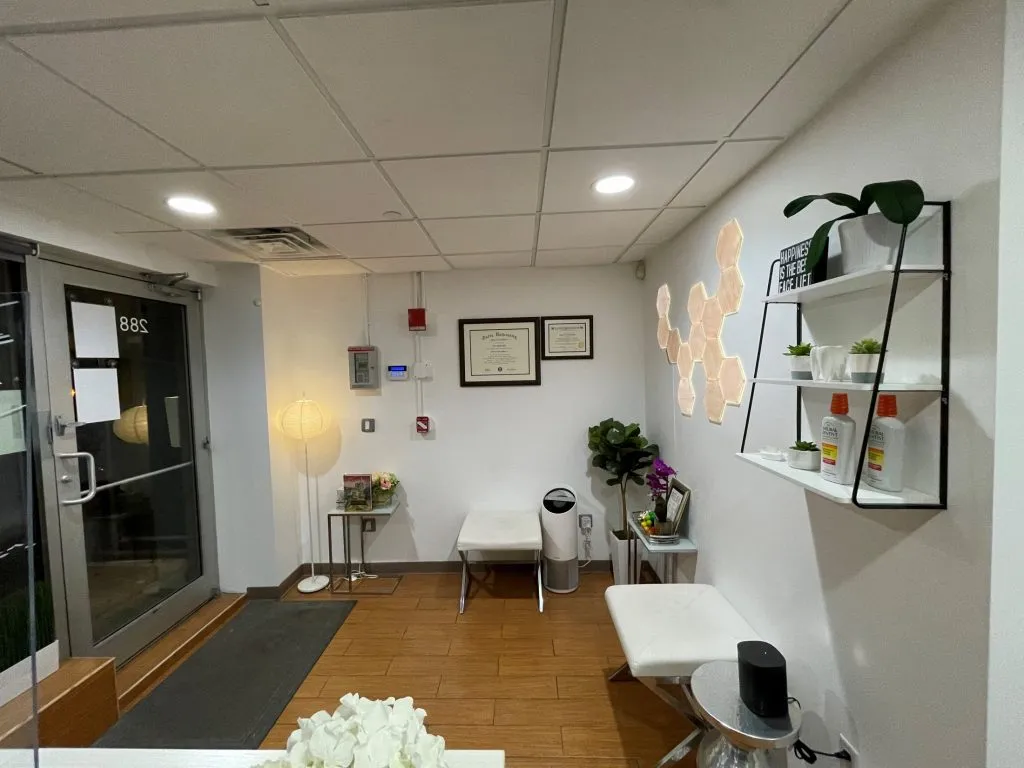
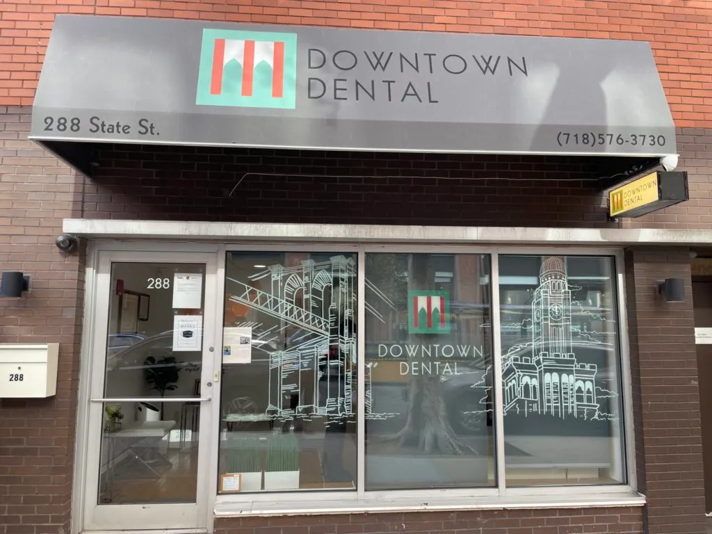
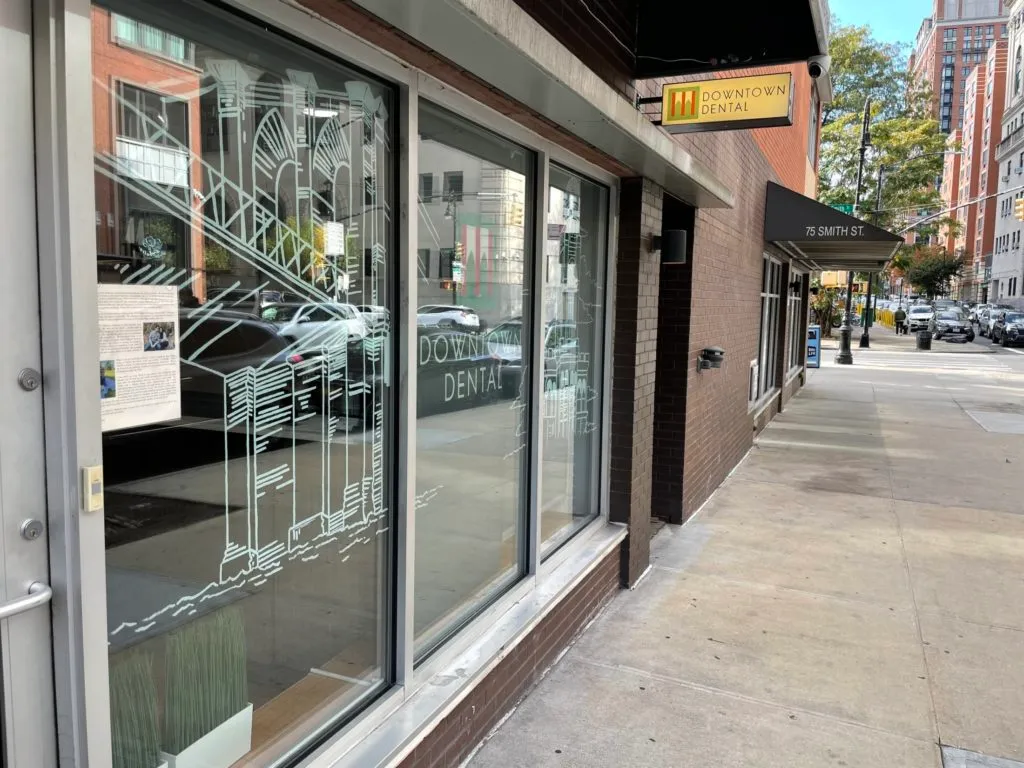
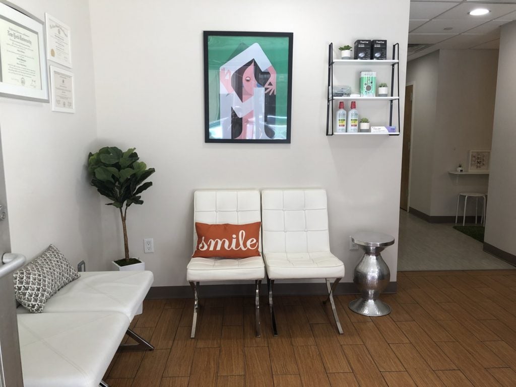
At Downtown Dental, your comfort and safety are our top priorities. We understand that visiting the dentist can sometimes be stressful, and that safety and cleanliness are important concerns. Whether you’re dealing with dental anxiety or just want reassurance about infection control, here’s what you can expect when you visit us in Downtown Brooklyn.
Overcoming Dental Anxiety & Phobia
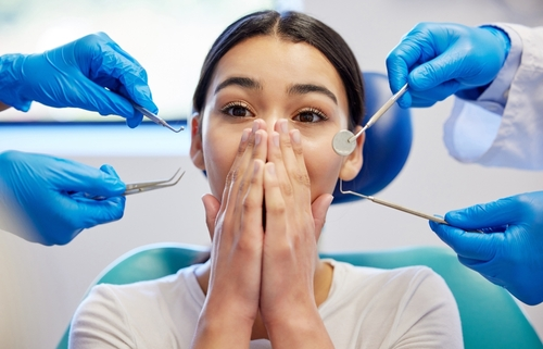
Many patients experience fear or anxiety about dental visits. You are not alone, and we are here to help. Our team takes extra care to make your visit as comfortable as possible:
-
Gentle communication and explanations about each procedure
-
Allowing breaks when needed during treatment
-
Calming environment with music and TV to distract and relax
-
Personalized approaches for nervous or anxious patients
Our goal is to make your visit stress-free while ensuring you receive the highest quality dental care.
Our Infection Control & Your Safety
Your safety is just as important as your comfort. Downtown Dental follows rigorous infection control procedures recommended by the CDC, OSHA, and EPA, including:
-
Hand hygiene and disinfecting surfaces
-
Wearing gloves and masks
-
Sterilizing all reusable instruments in an autoclave
-
Using disposable materials whenever possible
We are committed to maintaining a clean, safe, and trustworthy environment for every patient.
What to Expect During Your Visit
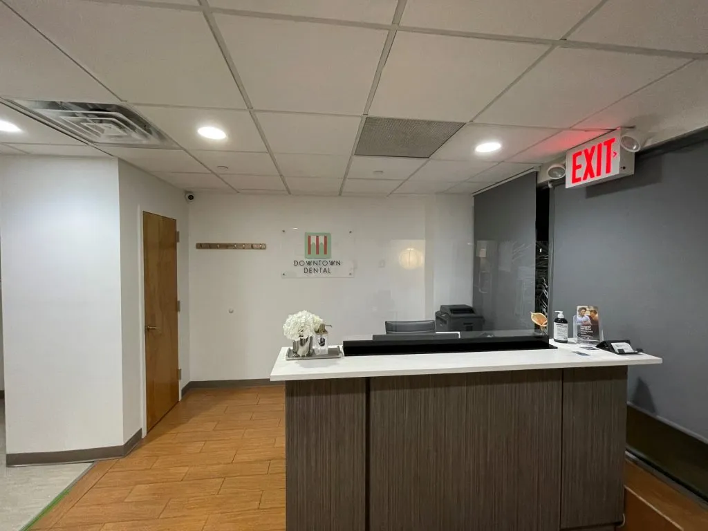
From the moment you walk in, you’ll notice our focus on comfort and safety. Our team is attentive to your concerns, whether it’s helping you feel relaxed or explaining our infection control practices. You can trust that your dental care in Downtown Brooklyn is both gentle and safe.
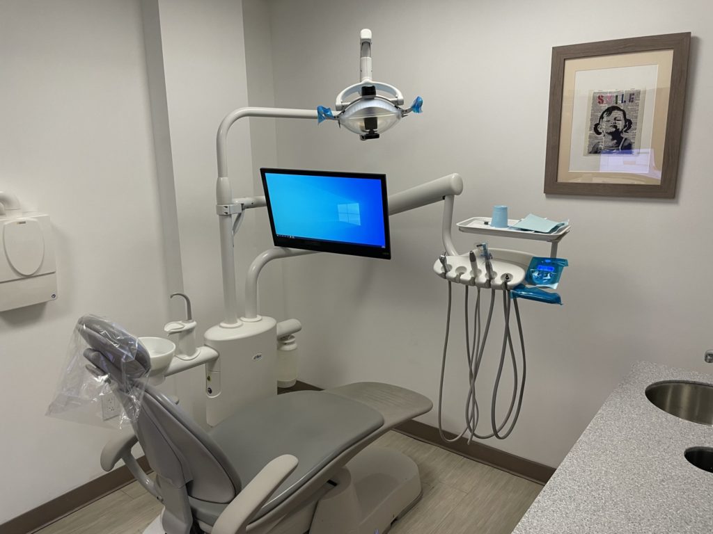
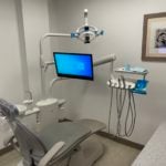
Advanced Dental Technology
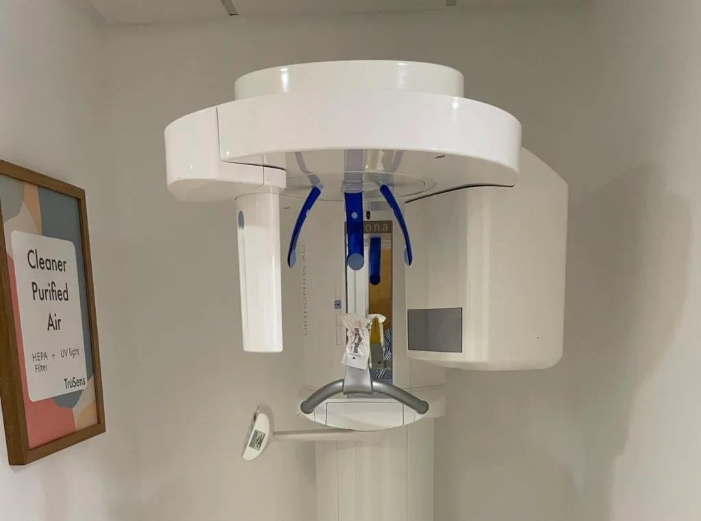
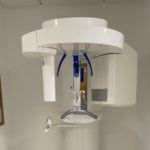
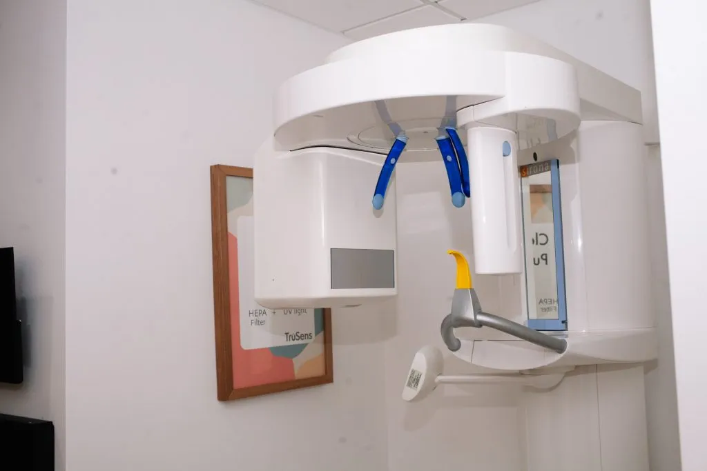
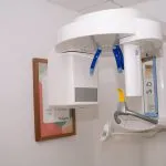
Panorex X-Ray
What is Panorex X-ray imaging?
Panorex X-ray imaging creates two-dimensional dental images displaying the upper and lower jaws and teeth. Panoramic X-rays are popular for dental applications because they are faster and less invasive than traditional X-ray imaging. They are used to detect, assess, and diagnose wisdom teeth impactions, periodontal disease, jaw health, and oral cancer.
What are the benefits of Panoramic dental imaging?
- Faster than traditional X-rays
- More comfortable for patients
- Easier for dentists to get the images they need
- Less exposure to radiation than traditional X-rays
- Higher diagnostic efficacy
- More hygienic and sanitary than traditional bitewing X-rays
How does a Panorex X-ray machine work?
Panorex X-ray digital imaging is extraoral, which means the X-ray machine does not have to go inside your mouth. Instead, the device works more like an MRI machine, rotating the X-ray tube around your head in a circle. The process is painless and only lasts between 10 to 20 seconds.
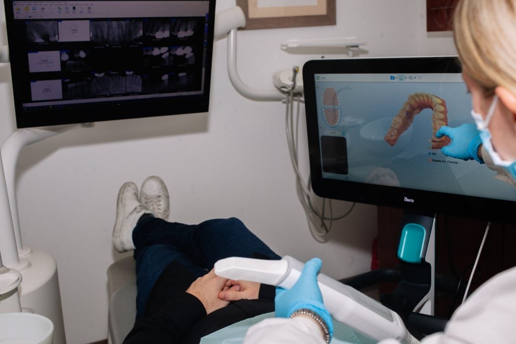
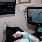
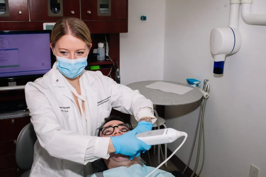
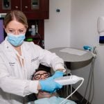
iTero® IntraOral Scanner
What is the iTero® IntraOral Scanner?
We are proud to offer the iTero® IntraOral Scanner to our patients for enhanced comfort and treatment outcomes. With the iTero® IntraOral Scanner, we can produce digital impressions with cost-saving benefits, and patients can see predicted results before treatment with instant 3D imaging – no goopy impression material required. Impressions with the iTero® IntraOral Scanner aren’t just more comfortable, they are also more accurate, giving our patients better results every day. Call us to find out more about the iTero® IntraOral Scanner today! Downtown Dental Office Phone NumberDowntown Dental Office Phone Number 718-576-3730
What are the benefits of iTero® IntraOral Scanner?
- Single appointment
- Comfortable
- Predictable and accurate
- Less chair time
- Digital records
- Chairside visualization and planning
How does the iTero® IntraOral Scanner work?
A compact, handheld wand is used to digitally map the structure of your teeth and gums using optical technology. In just 2-3 minutes, we have an accurate picture of your oral anatomy, allowing us to plan precision treatments that will yield the best results for you.
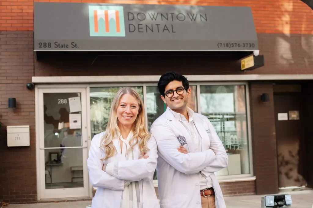
Precision Dentistry
When you seek care at our office, you are assured that Dr. Shah, Dr. Moses, and their team utilize the latest in technology to enhance the quality and fit for your dental care.
Digital Imaging
Drs. Shah and Moses choose carefully which and when radiographs are taken. There are many guidelines that we follow. Radiographs allow us to see everything we cannot see with our own eyes. Radiographs enable us to detect cavities in between your teeth, determine bone level, and analyze the health of your bone. We can also examine the roots and nerves of teeth, diagnose lesions such as cysts or tumors, as well as assess damage when trauma occurs.
Dental radiographs are invaluable aids in diagnosing, treating, and maintaining dental health. Exposure time for dental radiographs is extremely minimal. Drs. Shah and Moses utilize Digital Imaging Technologies within the office. With digital imaging, exposure time is about 50 percent less when compared to traditional radiographs. Digital imaging can also help us retrieve valuable diagnostic information. We may be able to see cavities better.
Digital imaging allows us to store patient images and enables us to quickly and easily transfer them to specialists or insurance companies.
Digital X-Rays:
Digital X-rays offer more precision since we view the image on a computer monitor, instead of holding up a 35mm film up to the light. Digital X-rays result in 1/6th of the radiation exposure to you.
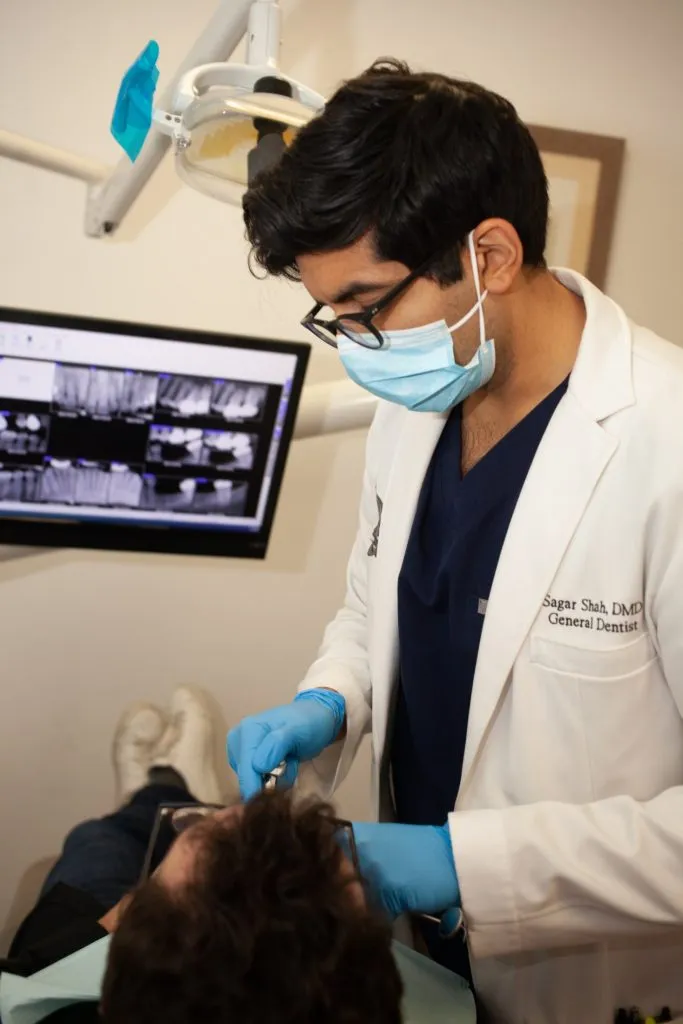
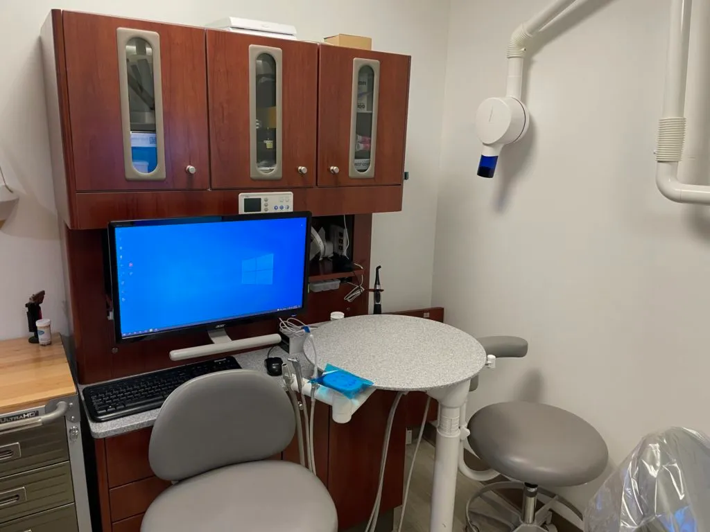
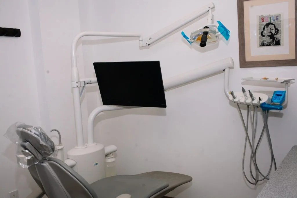
Intraoral Camera
Many patients, especially younger patients, are very familiar with the latest technology and are comfortable with the high-tech practice. Computers and TV screens are their primary method of information processing.
Dr. Shah and Dr. Moses utilize Intraoral Camera technology that helps enhance your understanding of your diagnosis. An Intraoral Camera is a very small camera – in some cases, just a few millimeters long. An Intraoral Camera allows our practice to view clear, precise images of your mouth, teeth, and gums, in order for us to accurately make a diagnosis. With clear, defined, enlarged images, you see details that may be missed by standard mirror examinations. This can mean faster diagnosis with less chair-time for you!
Intra oral cameras also enable our practice to save your images in our office computer to provide a permanent record of treatments. These images can be printed for you, other specialists, and your lab or insurance companies.
LED POWERED DENTAL UNITS
Our office is equipped with LED-powered dental units. This means all work that is done inside the oral cavity is illuminated with an LED-powered light.
LED-powered dental units provide optimal working environments making the patient comfortable and our work precise.